


 74 128

A
B
C
D
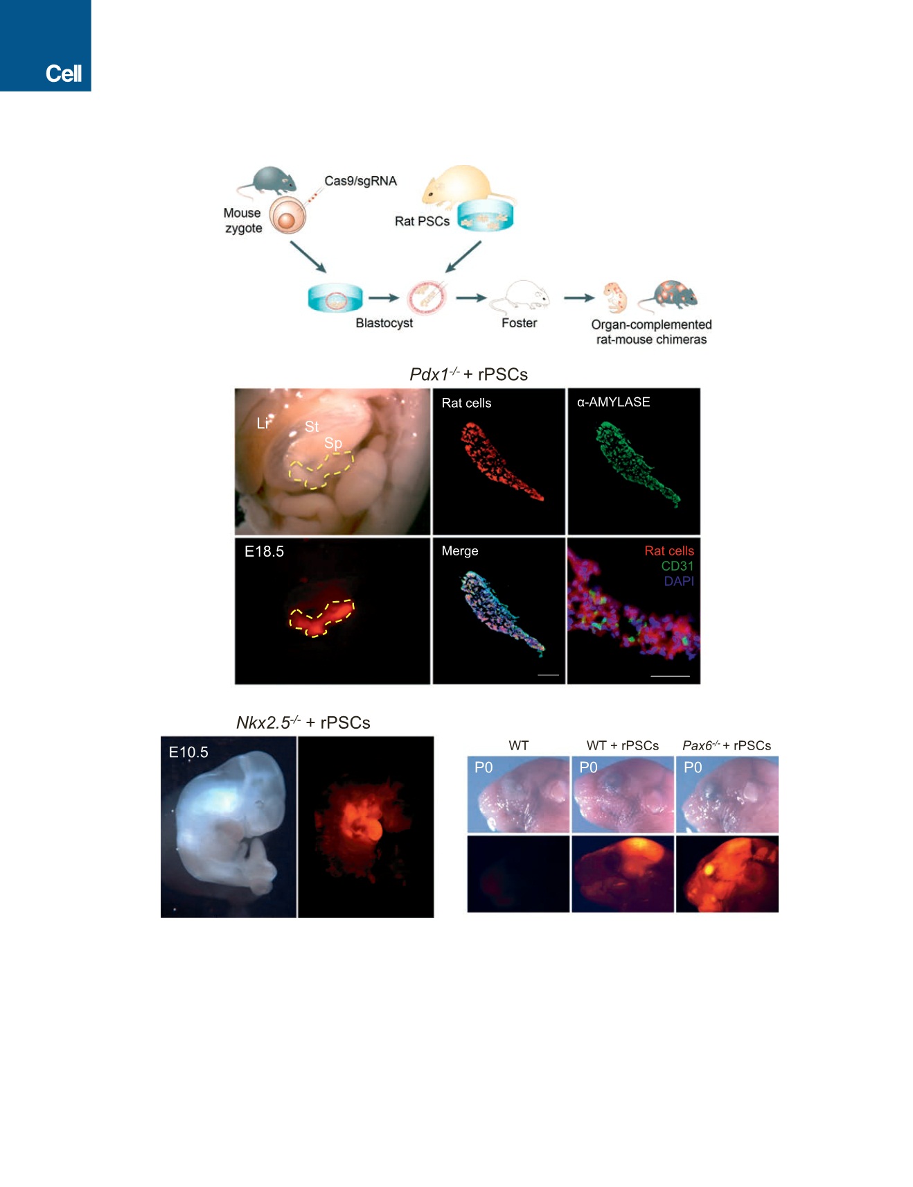
Figure 2. Interspecies Blastocyst Complementation via CRISPR-Cas9-Mediated Zygote Genome Editing
(A) Schematic of the CRISPR-Cas9 mediated rat-mouse blastocyst complementation strategy.
(B) Left, bright-field (top) and fluorescence (bottom) images showing the enrichment of rat cells in the pancreas of an E18.5
Pdx1
/
mouse. Li, liver; St, stomach;
Sp, spleen. Yellow-dotted line encircles the pancreas. Red, hKO-labeled rat cells. Middle and right (top), representative immunofluorescence images showing rat
cells expressed
a
-amylase in the
Pdx1
/
mouse pancreas. Blue, DAPI. Right (bottom), a representative immunofluorescence image showing that some
pancreatic endothelial cells, as marked by a CD31 antibody, were not derived from rat PSCs. Scale bar, 100
m
m.
(C) Bright field (left) and fluorescence (right) images showing the enrichment of rat cells in the heart of an E10.5 Nkx2.5
/
mouse. Red, hKO-labeled rat cells.
(D) Bright field (top) and fluorescence (bottom) images showing the enrichment of rat cells in the eye of a neonatal Pax6
/
mouse. Red, hKO-labeled rat cells. WT,
mouse control; WT+rPSCs, control rat-mouse chimera without Cas9/sgRNA injection.
See also Figure S2 and Tables S1 and S2.
476
Cell
168
, 473–486, January 26, 2017