


 46 128

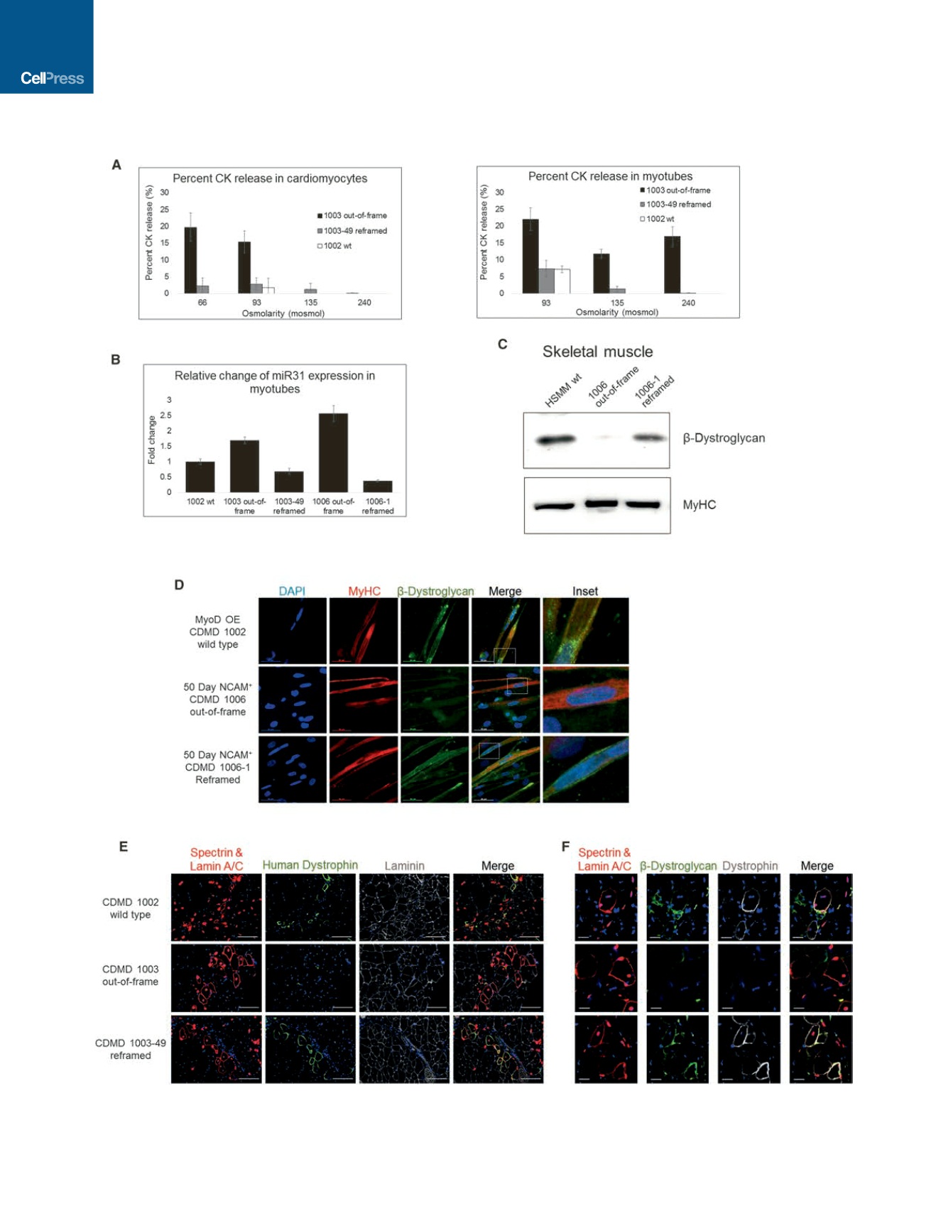
Figure 4. Reframed hiPSC-Derived Cardiomyocytes and Skeletal Muscle Cells Demonstrate Restored Function In Vitro and In Vivo
(A) Representative graphs of CK release assays from cells exposed to hypo-osmotic conditions. Cardiomyocytes and skeletal muscle myotubes derived from
hiPSCs were subjected to a range of osmolarities below 240 mosmol, and CK release to the supernatant was measured as an indication of membrane fragility.
Data are presented as average ± SE.
(legend continued on next page)
538
Cell Stem Cell
18
, 533–540, April 7, 2016
ª
2016 Elsevier Inc.